

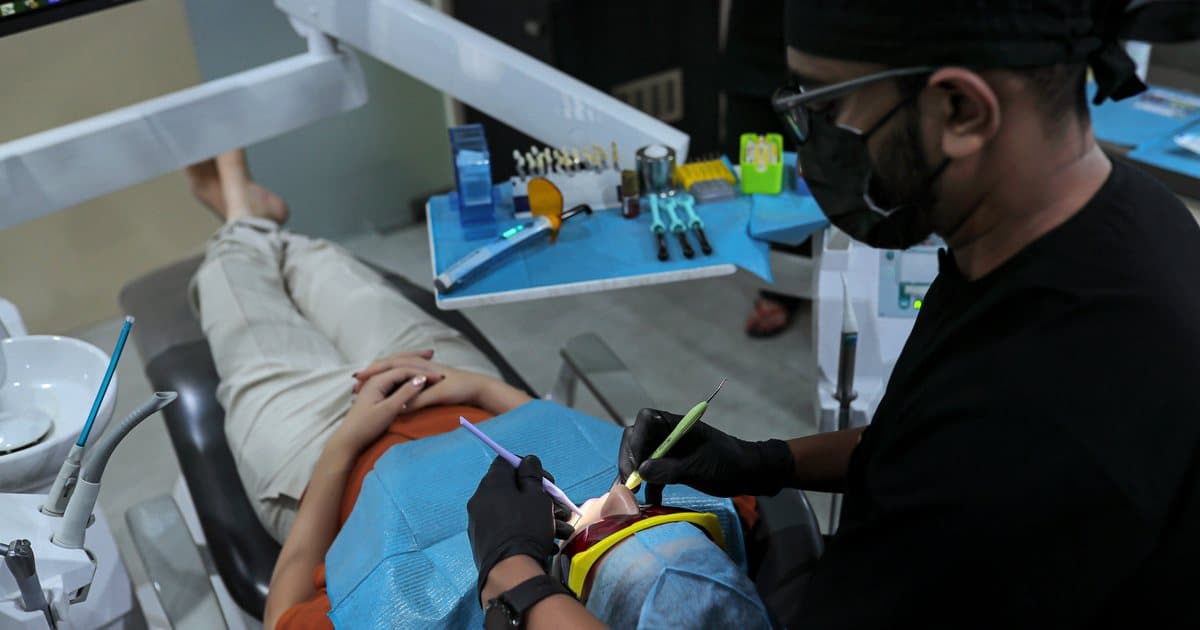
Dental Microscope in Dentistry – Why Precision Matters in Modern Root Canal Treatment
Modern dentistry is no longer based on approximation and limited visibility. Today, advanced treatments rely on precision, magnification, and technology to improve long-term success.
One of the biggest advancements in modern dental treatment is the Dental Operating Microscope.
This technology has completely transformed root canal treatment, restorative dentistry, and cosmetic procedures by allowing dentists to see details invisible to the naked eye.
This blog explains how dental microscopes work, why they matter, and why microscope-assisted dentistry is becoming the gold standard in modern dental care.
What Is a Dental Operating Microscope?
A dental microscope is a high-magnification device designed specifically for precision dental procedures.
It provides:
- Up to 25× magnification
- Powerful LED illumination
- Enhanced visibility of tiny structures inside the tooth
This allows dentists to detect and treat problems that traditional methods often miss.
Why Visibility Matters in Dentistry
In many dental procedures, success depends on identifying:
- Hidden canals
- Tiny cracks
- Residual infection
- Curved anatomy
- Microscopic leakage
These details are often impossible to see clearly with the naked eye.
The microscope improves:
- Diagnosis
- Accuracy
- Cleaning quality
- Long-term treatment success
How Dental Microscopes Improve Root Canal Treatment
Root canal treatment is one of the procedures that benefits most from microscope technology.
Microscopes Help Detect:
- Extra canals
- Calcified canals
- Micro fractures
- Incomplete cleaning
- Broken instruments inside canals
This significantly improves the quality of treatment.
Traditional RCT vs Microscopic RCT
Traditional Root Canal Treatment
- Limited visibility during treatment
- Higher chance of missed canals
- Average treatment precision
- Greater risk of re-infection
- Standard dental procedure
Microscopic Root Canal Treatment
- Up to 25× magnification for better visibility
- Improved detection of hidden canals
- Highly precise treatment approach
- Higher success rate with reduced re-infection
- Advanced precision-based dentistry
Advantages of Microscope Dentistry
1. Better Accuracy
Magnification allows precise treatment with minimal guesswork.
2. Conservative Dentistry
Only infected areas are treated, preserving more natural tooth structure.
3. Higher Success Rates
Microscopic precision reduces treatment failure and re-infection.
4. Faster Healing
Accurate treatment causes less trauma to surrounding tissues.
5. Improved Long-Term Results
Better cleaning and sealing improve tooth longevity.
Procedures That Benefit from Dental Microscope Technology
Dental microscopes are commonly used for:
- Root canal treatment
- Re-root canal procedures
- Broken file removal
- Cosmetic restorations
- Veneers and crown margins
- Crack detection
- Conservative fillings
Why Many Root Canals Fail
Most failed root canals happen because:
- Hidden canals were missed
- Infection remained inside the tooth
- Cracks were undetected
- Cleaning was incomplete
Microscope-assisted treatment reduces these risks significantly.
Dental Microscope and Digital Dentistry
Modern advanced clinics combine:
- Dental microscopes
- CAD-CAM systems
- Digital scanners
- 3D imaging
This combination creates:
- More predictable outcomes
- Better fitting restorations
- Improved patient comfort
Technology today is focused on precision-driven dentistry.
Is Microscope Dentistry Worth It?
Yes — especially for:
- Complex root canals
- Retreatment cases
- Long-term tooth preservation
- Patients wanting the best possible outcome
While microscope-assisted treatment may cost slightly more, it reduces:
- Repeat procedures
- Treatment failures
- Future complications
Long-term value matters more than short-term savings.
Common Patient Misconception
Many patients believe:
“All root canals are the same.”
They are not.
The difference between traditional and microscope-assisted treatment is similar to:
- Basic surgery vs microsurgery
- Manual precision vs magnified precision
Technology changes outcomes.
Final Thoughts
Dental microscopes are changing modern dentistry by making treatments:
- More accurate
- More conservative
- More predictable
- More durable
Microscope-assisted dentistry is no longer a luxury—it is becoming the standard for advanced dental care.
If long-term success matters, precision matters.
Frequently asked questions
What is a dental operating microscope?
A dental operating microscope is a magnifying device with bright, focused light that lets the dentist see fine details inside and around a tooth, such as extra canals, tiny cracks and the edges of fillings. It is used for root canals, re-treatment, broken instrument removal and precise restorations, instead of working with the naked eye.
Is microscopic root canal treatment better than normal RCT?
For hidden canals, cracks, calcified canals and re-treatment, a microscope helps because the dentist can see what the naked eye misses, and an untreated canal is a common reason root canals fail. The microscope removes guesswork but does not replace skill; the dentist's training and careful cleaning still matter most.
How much does microscopic root canal treatment cost in Surat?
At Ocean 32, microscopic root canal treatment typically costs ₹7,000 to ₹14,000 per tooth, and re-treatment of a failed root canal ₹8,000 to ₹16,000 per tooth. The fee depends on the tooth and its complexity and is confirmed in a written estimate after examination and X-rays. Back teeth usually also need a crown or onlay.
Can a broken instrument in a root canal be removed?
Usually, yes. Under the microscope, a file fragment left in a canal can often be located and removed using specialised tools such as the BTR-Pen, after which the canal is cleaned and sealed. Whether removal is possible depends on where the fragment sits and the shape of the root, which X-rays and examination reveal.
Who uses the dental microscope at Ocean 32?
Dr. Kenil A. Lathia, an MDS root canal specialist, has worked under the dental microscope since 2016 and uses a Labomed surgical microscope for every procedure: root canals, fillings, inlays, onlays, crowns and veneers. He is a Gold Member and moderator of the International Microscopic Dental Club, a microscope-dentistry community of dentists worldwide.